Ashley Makela
Assistant Professor
The University of Texas MD Anderson Cancer Center
Department of Imaging Physics
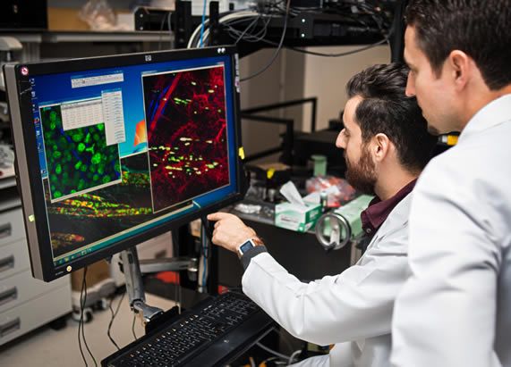
My lab uses molecular imaging to guide the development of therapeutic strategies for cancers that do not respond well to traditional treatments—such as those driven by cancer stem cells (CSCs), located in hypoxic niches, or metastasized to the brain. We integrate imaging, cancer biology, and drug delivery approaches to understand tumor heterogeneity, optimize therapeutic targeting, and assess treatment responses in real time.
Projects in the lab span several interconnected areas: (1) targeting CSCs in hypoxic tumor microenvironments using an engineered phosphatidylserine-targeting approach; (2) using extracellular vesicles (EVs) as delivery vehicles to reach and treat brain metastases; (3) applying imaging to monitor dynamic changes in the tumor microenvironment following therapy; and (4) tracking immune cell trafficking and activity when used as cancer therapeutics. Across these efforts, we utilize advanced imaging techniques—including bioluminescence, fluorescence, and magnetic particle imaging (MPI)—to visualize therapeutic delivery and efficacy in vivo.
Students rotating in the lab will gain hands-on experience with a range of techniques, including EV isolation and labeling, cell culture, flow cytometry, fluorescence microscopy, cell imaging assays, nanoparticle conjugation, and preclinical imaging in small animal models. Emphasis will be placed on experimental design, data analysis, and critical interpretation. Rotators will have the opportunity to contribute to translational research at the interface of drug development, nanomedicine, and tumor biology, with projects tailored to their interests and long-term goals.
Key words: cancer stem cells, brain metastasis, extracellular vesicles, tumor microenvironment, drug delivery, hypoxia, molecular imaging, bioluminescence, magnetic particle imaging, breast cancer
Education & Training
PhD - University of Western Ontario - 2019


