


Listing Not Found. Return to faculty directory.
 UTHealth - Houston's Health University
-
MS PROGRAMS
Thesis Based MS Programs
Specialized MS
Individualized MS Program in Biomedical Sciences
-
PHD PROGRAMS
PhD Programs
Overview
-
MD/PhD PROGRAM
MD/PhD Program
Participating Institutions/Entities
-
Admissions
Admissions
Admission FAQs
Admissions Office
6767 Bertner Avenue
S3.8344 Mitchell BSRB
Houston TX 77030 -
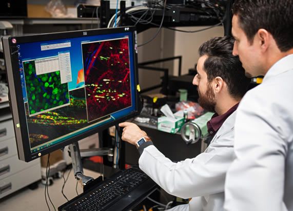
Research
Research
Research Interests
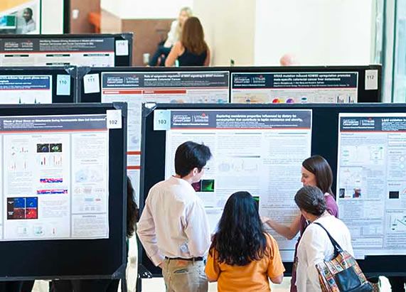
Student Research Day 2020
-
Student Life
Student Life
Student Organizations