


August’s Paper of the Month reveals how the RAS/ERK pathway orchestrates the opening notes of life
August 29, 2025
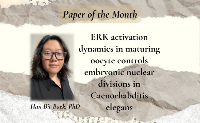


Paper of the Month: ERK activation dynamics in maturing oocyte controls embryonic nuclear divisions in Caenorhabditis elegans
Reproduction is among nature's most elegant orchestras. Diverse biomolecules and signaling partners perform in harmony to create a living organism. Despite each “movement” having a unique tempo, they converge beautifully. The orchestra’s opening sonata is gametogenesis. Haploid gametes (oocyte and sperm) with distinct genetic make-up are formed. Next is fertilization, where the gametes combine their genetic melodies to form a diploid zygote. During embryogenesis, the orchestra’s pace intensifies as the zygote divides to produce the multicellular embryo. The conclusion of this powerful symphony is life itself!
The maternal oocyte is the conductor of reproduction. It plays a pivotal role in setting the rhythm for embryogenesis. Further, the oocyte is the primary supplier of the genetic toolkit of mRNA, proteins, and signaling cues needed for embryonic development. Among these molecular players is the RAS/ERK pathway. This signaling cascade helps channel cues from the upstream director RAS (Rat Sarcoma Virus), a small GTPase, to turn on its effector enzyme, ERK (Extracellular Signal-Regulated Kinase). ERK is a terminal kinase that regulates oocyte maturation into an embryo. Previously, these researchers reported that across multiple species, ERK activity switches off during the oocyte-to-embryo transition. However, what would happen if ERK reactivated during this transition? To date, the answer remains elusive. This poses a critical gap in our understanding of ERK pathway dynamics in the embryo’s earliest stages of development.
In her doctoral research recently published in Cell Reports, Han Bit Baek, PhD, investigated the developmental mystery of why ERK signaling must be regulated with temporal precision during early embryogenesis. A recent graduate of the Genetics and Epigenetics program, Baek reflected on the experimental discovery that set her research in motion. “Pilot data suggested that ectopic ERK activation prevented oocyte and sperm pronuclei fusion and subsequently hindered zygotic nucleus formation,” she observed. Inspired by these results, she was keen to interrogate the consequences of abnormal ERK activity and how it might reshape the developmental score of embryonic development.
When the first notes fall out of tune: abnormal ERK activation during oocyte maturation
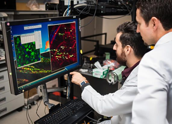
Baek and the article's coauthors in the research group of Swathi Arur, PhD, used C. elegans as the model system to study the effects of aberrant ERK signaling on oocyte maturation. “C. elegans and humans share many of the same fundamental biological pathways,” Baek explains. The nematode’s transparent body allows scientists to watch molecular processes unfold in real time through live imaging. Furthermore, these microscopic roundworms are easy to grow and maintain in the laboratory, reproduce quickly, and are amenable to genetic editing. For Baek, C. elegans was not just a model system, but an ideal stage to watch the symphony of oocyte-to-embryo transition play out at the molecular level.
Baek and the co-authors employed RAS(act), a previously described RAS-gain-of-function mutant, to ectopically activate ERK expression in the C. elegans embryos. ERK hyperactivation altered oocyte maturation and yielded defective embryos. Abnormalities in morphology, chromosome dynamics, and mitotic nuclear divisions characterized these embryos. Through confocal and live cell imaging, Baek also learned that the oocyte and sperm pronuclei had failed to fuse. Instead, the embryos exhibited a distinctive “paired nuclei” phenotype characterized by the blastomere containing two separate nuclei, maternal and paternal. This unique defect provided Baek with the first clue that aberrant ERK activation prevents embryonic nuclear envelope breakdown (NEBD) from occurring and sabotages the developmental progression of the embryo.
Apotheosis: ERK orchestrates the inhibition of PLK-1
Eager to uncover how abnormal ERK activation blocks NEBD, the authors interrogated the molecular basis of the paired nuclei phenotype. Previous work in the field demonstrated that depleting Polo-like kinase 1 (PLK-1) yielded the paired nuclei phenotype she observed in RAS(act) embryos. Using in vitro kinase assays, Baek learned that ERK inhibits PLK-1 by phosphorylating a canonical site — S404. The next piece of the puzzle that Baek and her team were eager to uncover was the molecular mechanism disrupting PLK-1 function in RAS(act) mutant C. elegans embryos. “I was keen to test whether the effects of aberrant ERK activation affected PLK-1 localization and abundance,” she mused.
After crossing in a partial loss-of-function allele, plk-1(rf) (GFP-tagged PLK-1), into RAS(act) animals, Baek discovered that PLK-1 localization was unaffected. Upon comparing PLK-1 protein levels in plk-1(rf) and plk-1(rf); RAS(act) animals, Baek saw no change in PLK-1 abundance. Based on these findings, she determined that RAS(act) neither perturbed PLK-1 localization or abundance.
However, while performing these experiments, Baek made a breakthrough discovery. She observed that introducing the RAS(act) mutation in plk-1(rf) animals increased penetrance of the paired nuclear phenotype. “These results were exciting since they suggest that ERK and PLK-1 functionally intersect to regulate NEBD in the early embryo,” she hypothesized. Her hunch proved true when removal of the ERK-dependent phosphorylation site on PLK-1 rescued the nuclear defects in plk-1(rf); RAS(act) double mutant embryos. Like a melody reversed, the converse also held. When the ERK-dependent PLK-1 phosphorylation site was constitutively phosphorylated, the paired nuclear defect was significantly enhanced. Jubilant, Baek concluded that abnormal RAS/ERK activation mediated PLK-1 phosphorylation and subsequent loss of NEBD between the oocyte and sperm pronuclei.
Connecting the dots in the score of oocyte-to-embryo transition
These results illuminate the previously unresolved mystery of how RAS/ERK signaling modulates PLK-1 to control nuclear dynamics. “Our findings reveal a new layer of developmental regulation and suggest broader intersections between these pathways,” Baek said. By tying the RAS/ERK pathway to PLK-1 activity, Baek showed that developmental success depends not just on whether key pathways are switched on, but also on the precise moment they are turned off. Reflecting on the work, she described the discovery as “connecting the dots of a non-linear puzzle,” where each surprising piece eventually snapped into place to reveal a coherent picture of the cellular and molecular mechanisms governing early embryonic development.
Since ERK and PLK-1 proteins are evolutionarily conserved, Baek speculates that this signaling axis could play a pivotal role in human embryonic development. “Alterations in this signaling axis could affect the success rate of assisted reproductive technologies like in vitro fertilization (IVF),” Baek thinks. In IVF, hormonal stimulation is commonly used to perform superovulation. However, such techniques increase the likelihood of ectopic ERK activation through the cyclic AMP (cAMP)–protein kinase A (PKA) pathway. This phenomenon could affect oogenesis, leading to an oocyte that might not be developmentally competent to support healthy embryogenesis. Thus, developing noninvasive methods and biomarkers that report abnormal ERK activity may dramatically improve IVF outcomes.
The most striking lesson one can glean from Baek’s research is the power of timing in biology. As in music, where a single instrument out of sync can throw off the entire performance, in development, a pathway activated at the wrong moment can derail the concert of creation. For Baek, her research journey went beyond collecting data. It became an exercise in learning to read the hidden score of cellular life. “Writing this article allowed me to synthesize my data and truly appreciate its biological significance,” she reflected. By showing that ERK needs to quiet down for the concert of life to progress uninterrupted, Baek has brought forth a new movement in the symphony of embryonic development that can reshape future research and clinical practice.
Baek had the opportunity to present her work, along with other Graduate School students at an MD Anderson Research Town Hall earlier this year. Congratulations on earning her PhD in May, we look forward to following her journey as an alum!

Arur Lab day out at Houston Rodeo 2025!
Paper of the Month (POM) is a collaborative effort led by Microbiology and Infectious Diseases PhD candidate Jana Gomez and Communications Manager Shelli Manning, and overseen by Associate Dean for Academic Affairs Francesca Cole, PhD, who collaborate with students to summarize fellow student-authored scientific articles about their biomedical science research and the innovative methods and discoveries they are uncovering. The POM editorial team includes students Shraddha Subramanian (author of August’s POM summary), Mirrah Bashir, Amanda Warner, Chae Yun Cho, Altai Enkhbayar, Zarmeen Khan, and Archit Gupta.