


David Piwnica-Worms
Professor
The University of Texas MD Anderson Cancer Center
Departments of Cancer Systems Imaging and Cancer Biology
Genomic lesions within incipient cancer cells in collaboration with alterations in the microenvironment contribute to neoplastic progression. Tumor cells can modulate the surrounding microenvironment to promote the progression of cancer through intrinsic oncogenic pathways. However, the importance of the host microenvironment in neoplastic progression, independent of tumor manipulation, is also underscored by studies demonstrating that many stromal and immune cell types stimulate growth of pre-neoplastic and neoplastic cells along with promoting drug resistance. Given these observations, understanding the complex interactions between genomic lesions and tumor microenvironment in animal models is crucial to understanding mechanisms of transformation and uncovering new anti-cancer therapies. Thus, non-invasive imaging technologies have become increasingly important for providing spatial and temporal resolution of biological structure and function, particularly for defining the context of gene expression and protein function, and their regulatory mechanisms within the proper physiologic context of cellular micro-environments. Molecular imaging is used to interrogate protein processing, protein-protein interactions, gene expression and flux through metabolic pathways in real-time in cells, live animals, and humans, and is an increasingly useful tool for understanding signal transduction, pharmacodynamics, and the pathobiology of human diseases in vivo, facilitating development of effective therapies.
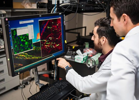
Techniques: Molecular imaging is broadly defined as the characterization and measurement of biological processes in living animals, model systems and humans at the molecular and cellular level using remote imaging detectors such as PET, SPECT, hyperpolarized MR, bioluminescence, and near-infrared fluorescence. We actively incorporate single cell imaging strategies, high throughput screens of cell phenotypes, cell and molecular biology techniques and conjugation chemistry into our projects.
Education & Training
M.D., Ph.D. - Duke University - 1984